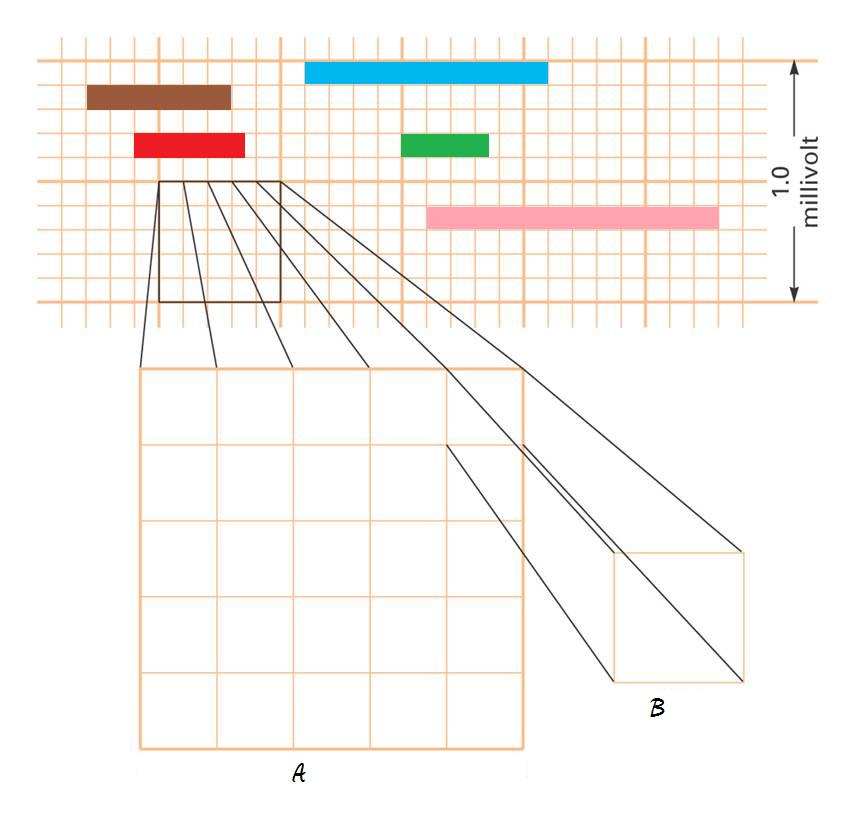
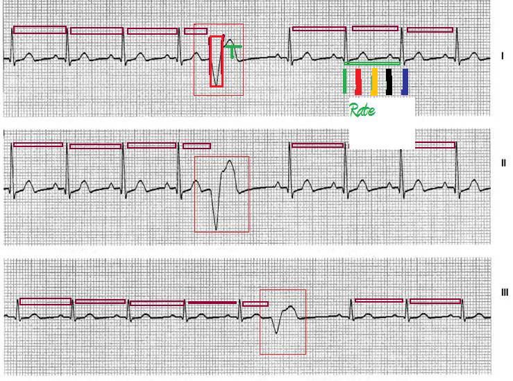
The letter A represents how much
time in seconds?
The letter B represents how much time in seconds?
For the following colors calculate the time in seconds?
The Brown line in seconds?
The Blue line in seconds?
The Green line in seconds?
The Red line in seconds?
The Pink line in seconds?
Using the triplicate method of calculating rate (large boxes into 300) what is
the rate for:
The Brown line?
The Blue line?
The Green line?
The Red line?
The Pink line?
Using the most accurate method to calculate rate (divide small boxes into
1500) what is the rate for:
The Brown line?
The Blue line?
The Green line?
The Red line?
The Pink line?
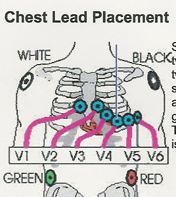
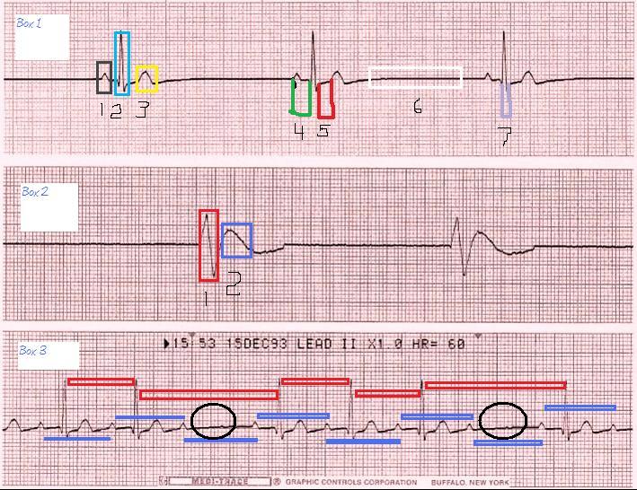
Using box 1: Black square numbered
1 is the?
Using box 1: Blue square numbered 2 is the?
Using box 1: Yellow square numbered 3 is the?
Using box 1: The green gap numbered 4 is the?
Using box 1: The red gap numbered 5 is the?
Using box 1: The white box numbered 6 is termed the?
Using box 2: The red rectangle represents the?
Using box 2: The blue rectangle represents the?
Using box 3: What is missing in the Black Circles?
Using box 3: Is the rhythm regular or irregular?
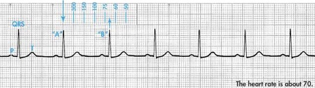
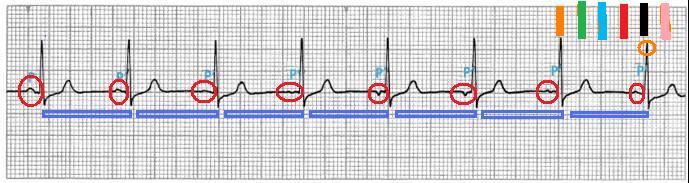
The P waves, which are circled RED,
are they the same or different?
Is the rhythm regular or irregular?
Using the QRS, which tip is circle in orange, what is the rate if it falls on
the green line using the triplicate method?
Using the QRS, which tip is circle in orange, what is the rate if it falls on
the red line using the triplicate method?
Using the QRS, which tip is circle in orange, what is the rate if it falls on
the blue line using the triplicate method?
Using the QRS, which tip is circle in orange, what is the rate if it falls on
the black line using the triplicate method?
Using the QRS, which tip is circle in orange, what is the rate if it falls on
the pink line using the triplicate method?
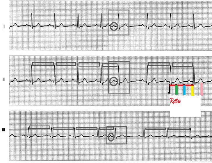
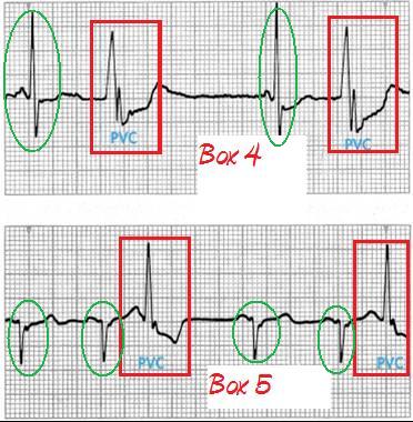
Premature beats are ectopic beats.
To be a premature beat it must come before the next expected normal beat. In
the square boxes are premature beats. There are three premature beats which
are PVC, PAC, PJC. The key difference is the origin of the beat. If there is
an upright P wave it is a PAC. If the P wave is absent or inverted and the
QRS is narrow is it a PJC. If the QRS is wide it is a PVC. The circle is the
area to focus on to determine which premature beat it is.
Is the premature beat a:
Is the rhythm regular or irregular? (You do not count the premature beat complex)
What is the rate if it falls on the green line using the triplicate method?
What is the rate if it falls on the pink line using the triplicate method?
What is the rate if it falls on the blue line using the triplicate method?
What is the rate if it falls on the yellow line using the triplicate method?
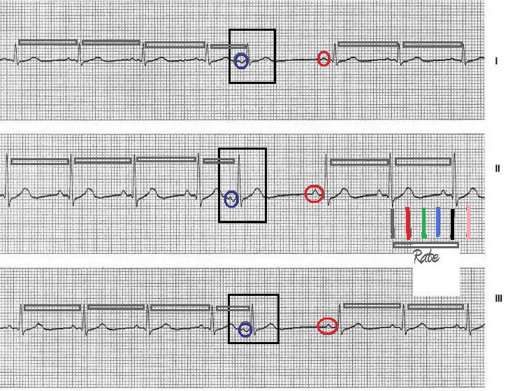
Premature beats are ectopic beats.
To be a premature beat it must come before the next expected normal beat. In
the square boxes are premature beats. There are three premature beats which
are PVC, PAC, PJC. The key difference is the origin of the beat. If there is
an upright P wave it is a PAC. If the P wave is absent or inverted and the
QRS is narrow is it a PJC. If the QRS is wide it is a PVC. The circle is the
area to focus on to determine which premature beat it is.
Is the premature beat a:
Is the rhythm regular or irregular? (You do not count the premature beat complex)
What is the rate if it falls on the green line using the triplicate method?
What is the rate if it falls on the red line using the triplicate method?
What is the rate if it falls on the blue line using the triplicate method?
What is the rate if it falls on the pink line using the triplicate method?
Premature beats are ectopic beats.
To be a premature beat it must come before the next expected normal beat. In
the square boxes are premature beats. There are three premature beats which
are PVC, PAC, PJC. The key difference is the origin of the beat. If there is
an upright P wave it is a PAC. If the P wave is absent or inverted and the
QRS is narrow is it a PJC. If the QRS is wide it is a PVC. The circle is the
area to focus on to determine which premature beat it is.
Is the premature beat a:
Is the rhythm regular or irregular? (You do not count the premature beat complex)
What is the rate if it falls on the black line using the triplicate method?
What is the rate if it falls on the red line using the triplicate method?
What is the rate if it falls on the blue line using the triplicate method?
What is the rate if it falls on the yellow line using the triplicate method?
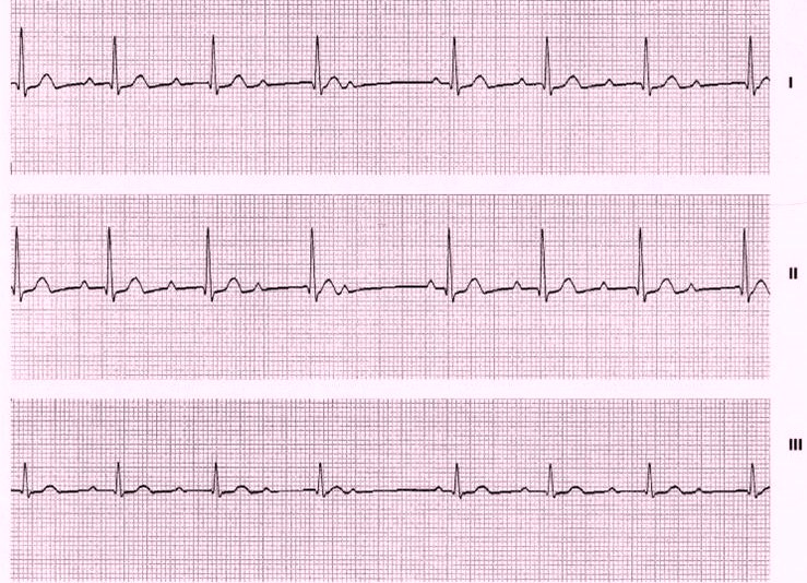
Is the QRS complete narrow or wide?
Is the rhythm regular or irregular?
What method must be used to calculate the rate?
Does every P wave have a QRS?
Does every QRS have a P wave?
Does the PR Interval stay the same measurement?
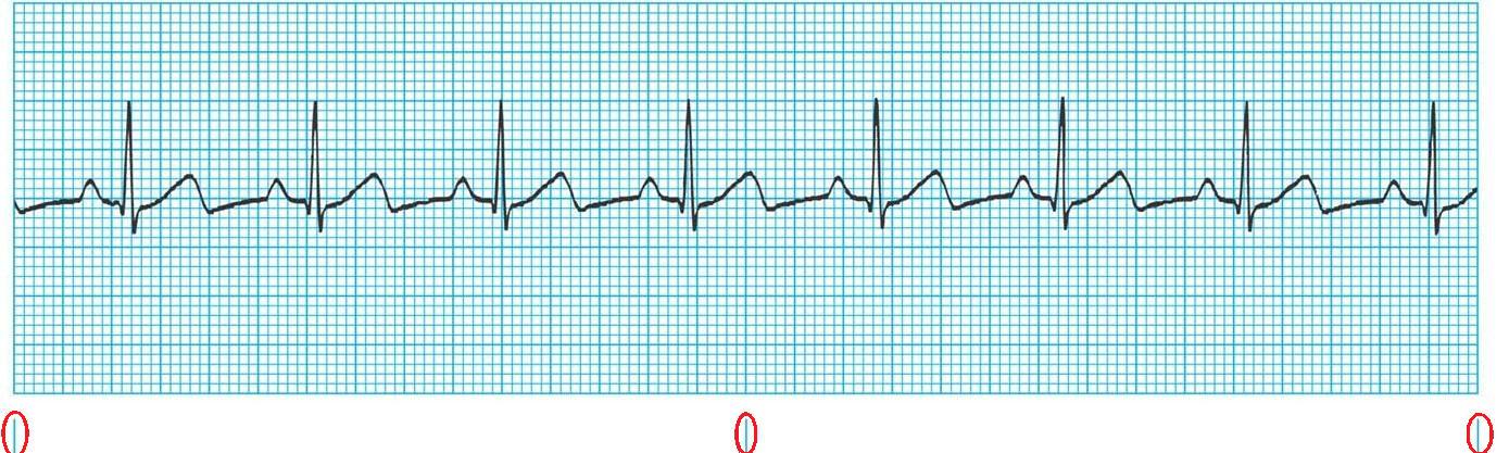
Using the ECG below answer the
following questions:
Is the rhythm regular or irregular?
Describe the P wave?
What is the PR Interval measurement?
What is the QRS measurement?
Is there one P wave for every QRS?
Is there one QRS for every P wave?
Is the T wave upright or inverted?
Using the 6 sec strip method to calculate rate what is the ventricular rate?
Using the most accurate way to calculate rate (little boxes divided into 1500)
what is the atrial rate?
Using the most accurate way to calculate rate (little boxes divided into 1500)
what is the ventricular rate?